
ТHIFEM technology for the treatment of urinary incontinence - mechanism of action
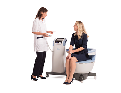
HIFEM (HIGH INTENSITY FOCUSED ELECTROMAGNETIC FIELD) TECHNOLOGY FOR THE TREATMENT OF URINARY INCONTINENCE - MECHANISM OF ACTION
HIFEM* technology causes deep stimulation of the pelvic floor muscles and restoration of neuromuscular control.
The effectiveness is mainly due to the impact of focused electromagnetic energy, penetration into the depth and stimulation of the entire pelvic floor area.
During each HIFEM session, thousands of supramaximal pelvic floor muscle contractions occur, which is very important for muscle retraining in incontinent patients.
Incontinent patients are unable to perform repetitive contractions due to weakened pelvic floor muscles.
The role of the pelvic floor muscles
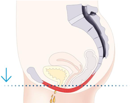
The pelvic floor muscles (PFM) are a group of muscles that support the organs of the pelvic floor and are responsible for holding urine. Due to the normal aging process, childbirth and menopause, the pelvic floor muscles are unable to properly support the pelvic floor organs. These conditions are directly related to incontinence.
Figure 1. Causes and consequences of urinary incontinence
Urinary incontinence
Urinary incontinence is defined as the involuntary leakage of urine. The International Society for the Diagnosis and Treatment of Urinary Incontinence distinguishes 3 main types of incontinence according to etiology:
Stress urinary incontinence (SUI) refers to the leakage of urine in situations where there is an increase in intra-abdominal pressure (including during coughing, sneezing, laughing, heavy lifting, etc.). SUI is caused by incompetence of the urethral sphincter and weakening of the pelvic floor muscles due to damage to the structures that support the pelvic floor. Also, the most common causes of stress urinary incontinence are childbirth and menopause.
The second type is associated with the occurrence of a strong urge to urinate and with pathological contractions of the bladder, this is the so-called urgency incontinence. Urge incontinence is a dysfunction of the neuromuscular system, usually a symptom of a deeper problem (including diabetes).
The third type is mixed urinary incontinence (MUI), which is a combination of the symptoms of the first two types - SUI and urge incontinence. In all three types, patients are unable to contract their pelvic floor muscles properly due to weakness in the pelvic floor muscles, in the case of SUI, or due to an overactive bladder, in the case of urge incontinence.
HIFEM Technology
HIFEM technology contributes to the creation of intense contractions of the pelvic floor muscles, acting on the neuromuscular apparatus and creating an electrical current in the tissues. Electric currents depolarize neurons, which leads to concentric contractions and elevation of all muscle groups of the pelvic floor. The effectiveness is mainly due to the deep penetration of focused electromagnetic energy and stimulation of the entire pelvic floor area. This contributes to a change in muscle structure, causing active growth of myofibrils (hypertrophy). New muscle fibers are synthesized - their hyperplasia occurs. HIFEM technology promotes deep stimulation of the pelvic floor muscles and restoration of neuromuscular control.
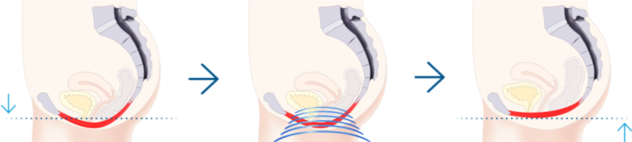
Figure 2. Comparison of the condition of patients before and after stimulation of the pelvic floor muscles using HIFEM technology
Supramaximal contractions of the pelvic floor muscles
Maximal voluntary contraction (MVC) is the greatest amount of tension that a muscle can generate and physiologically maintain, usually within a fraction of a second. Contractions with tension higher than MVC are called supramaximal. HIFEM technology helps create supramaximal pelvic floor muscle contractions and hold them for a couple of seconds (see Figure 1). These contractions are created independently of brain activity and are functionally associated only with the peripheral nerves of the pelvic floor area.
Supramaximal contractions cannot be achieved by the muscles voluntarily (including during Kegel exercises). The effectiveness of HIFEM technology is primarily due to the gradual increase in the intensity of the focused electromagnetic field and the frequency of the pulses, which gives a unique effect of creating energetic contractions. During the first HIFEM session, thousands of supramaximal contractions of the pelvic floor muscles are produced. This method is very important for muscle retraining, as patients are unable to perform these frequently repeated contractions due to weakened pelvic floor muscles.
This effect cannot be achieved with conventional exercises (including Kegel).
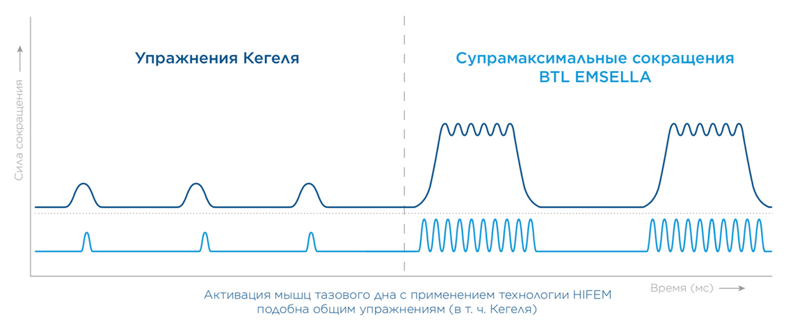
Figure 3. Pelvic floor muscle activation using HIFEM technology Time (ms) is similar to general exercises (including Kegel)
HIFEM Therapeutic Protocol
The HIFEM therapy protocol lasts 30 minutes and consists of 3 different phases. During these phases, the pelvic floor muscles are excited, then stimulated and relaxed. The repetition of these phases and exposure to focused electromagnetic energy leads to stimulation of the pelvic floor muscles, adaptation and remodeling.
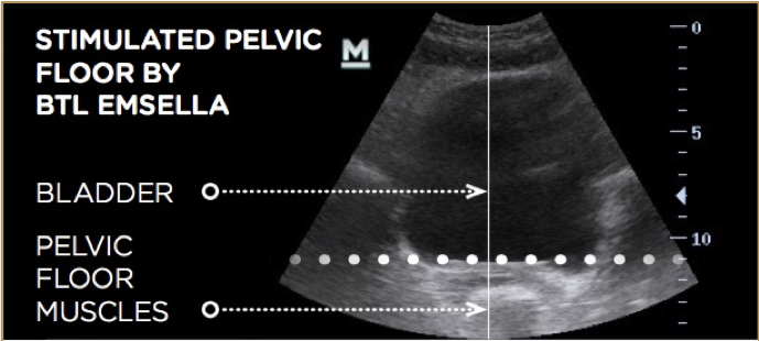
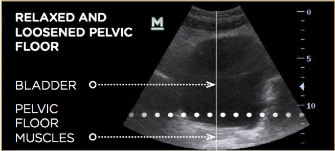
Figure 4. Ultrasound-based elevation of the pelvic floor muscles and bladder. Weakened pelvic floor muscles and bladder (right). Stimulated and elevated pelvic floor muscles and bladder after HIFEM technology (left)
Bibliography:
1. Abrams P, Blaivas JG, Stanton SL, Andersen JT. The Standardisation of Terminology of Lower Urinary Tract Function. The International Continence Society Committee on Standartisation of Terminology. Scand d Suppl 1998;114:5-19.
2. Almeida FG, Bruschini H, Srougi M.: Urodynamic and clinical evaluation of 91 female patients with urinary incontinence treated with perineal magnetic stimulation: 1-year follow-up. J Urol. 2004 Apr; 171(4), pages 1571-4.
3. Bickford, R., Guidi, M., Fortesque, P. and Swenson, M. (1987). Magnetic stimulation of human peripheral nerve and brain. Neurosurgery, 20(1), pp.110-116.
4. Coletti, D., Teodori, L., Albertini, M., Rocchi, M., Pristerà, A., Fini, M., Molinaro, M. and Adamo, S. (2007). Static magnetic fields enhance skeletal muscle differentiation in vitro by improving myoblast alignment. Cytometry Part A, 71A(10), pp.846-856.
5. Ishikawa N., Suda S., Sasaki T. et al., Development of a non-invasive treatment system for urinary incontinence using a functional continuous magnetic stimulator (FCMS) , Medical & Biological Engineering & Computing, 1998, 36, 704-71.
6. Ostrovidov, S., Hosseini, V., Ahadian, S., Fujie, T., Parthiban, S., Ramalingam, M., Bae, H., Kaji, H. and Khademhosseini, A. (2014). Skeletal Muscle Tissue Engineering: Methods to Form Skeletal Myotubes and Their Applications. Tissue Engineering Part B: Reviews, 20(5), pp.403-436.
7. Stölting, M., Arnold, A., Haralampieva, D., Handschin, C., Sulser, T. and Eberli, D. (2016). Magnetic stimulation supports muscle and nerve regeneration after trauma in mice. Muscle & Nerve, 53(4), pp.598-607.
8. Wallis, M., Davies, E., Thalib, L. and Griffiths, S. (2011). Pelvic Static Magnetic Stimulation to Control Urinary Incontinence in Older Women: A Randomized Controlled Trial. Clinical Medicine & Research, 10(1), pp.7-14.
You can subscribe and recive our weekly special offer right now